Min Lu1,2,
Xuan Zhou3,
Cheng-Guo Zheng2,
Feng-Jun Liu1 ![]() ,
,
For correspondence:- Feng-Jun Liu Email: liufengjun09@hotmail.com Tel:+8653182169114
Received: 4 April 2016 Accepted: 9 November 2016 Published: 20 December 2016
Citation: Lu M, Zhou X, Zheng C, Liu F, Expression profiling of miR-96, miR-584 and miR-422a in colon cancer and their potential involvement in colon cancer pathogenesis. Trop J Pharm Res 2016; 15(12):2535-2542 doi: 10.4314/tjpr.v15i12.1
© 2016 The authors.
This is an Open Access article that uses a funding model which does not charge readers or their institutions for access and distributed under the terms of the Creative Commons Attribution License (http://creativecommons.org/licenses/by/4.0) and the Budapest Open Access Initiative (http://www.budapestopenaccessinitiative.org/read), which permit unrestricted use, distribution, and reproduction in any medium, provided the original work is properly credited..
Purpose: To determine the correlation between miRNAs; miR-96, miR-422a and miR584, and colon cancer, and also to test whether any of these miRNAs can act as non-invasive biomarkers in colon cancer
Methods: The tumor samples and the corresponding normal mucosa used in this study were collected from 60 patients diagnosed with colon cancer. HT-29, HCT-116 and SW-620 cell lines were used for miRNA ex
Results: The results indicate down-regulation of miR-96, miR-584 and miR-422a in colon cancer tissue and decreased ex
Conclusion: This study shows that miR-96, miR-584 and miR-422a are down-regulated in colon cancer and are associated with tumor size. Thus, the ratio of miR-96/miR-638 in plasma is a potential non-invasive biomarker in colon cancer.
Introduction
MicroRNAs (miRNAs) are relatively newly discovered class of molecules that play crucial roles in cellular processes in humans and other mammals. miRNAs are transcribed by RNA polymerase II and do not code for any proteins, and are of varying lengths ranging between 20 and 25 nucleotides [1]. Previously unknown, miRNAs have been shown to play significant roles in cancer related pathways such as invasion, migration, proliferation, apoptosis and other aspects of tumorigenesis [2,3]. The precise mechanisms of miRNA function in mammals are still debatable. However, several years of literary evidence suggest that miRNAs regulate gene expression through precise sequence specific base pairing at the 3’ UTR of the target mRNA or at the translational level [4].
Colon cancer is the cancer of the colon with a million new cases reported each year globally [5]. In the United States alone, colon cancer is the third most prevalent form of cancer with 95,000 cases reported each year, and second most leading cause of cancer related deaths. Similar to other types of cancer, survival rates of colon cancer patients depend on the early detection of colon cancer. Therefore, it is important to detect colon cancer at an early stage. However, only 40 % of colon cancers are detected at an early stage. Several studies have shown that early detection of colon cancer is highly curable. The present methods of treatment include independent or a combination of surgical procedures, chemotherapy, radiotherapy and other targeted therapies depending on the stage of cancer. Since the afore-stated procedures are based on the late onset of colon cancer, it is important to develop early stage detection methods that have the potential to complement the aforementioned standard procedures to cure colon cancer or improve the life span of an individual.
Given the crucial association between miRNAs and cancer, several studies have classified different types of human cancers based on miRNA expression profiles [6,7]. Aberrant expression of miRNAs has been associated with almost every type of cancer. Expression analysis by RT-PCR revealed miR-221, miR-376a and miR-301 to be differentially expressed in pancreatic cancer providing new insights into pancreatic tumorigenesis [8]. A recent study has shown the let-7 miRNA cluster, miR-214, miR-199a*, miR-200a and miR-100 to be frequently deregulated in ovarian cancer [9]. Similarly, miRNA expression profiling of prostate and breast cancer cells have revealed a total of 51 miRNAs that were either upregulated or downregulated in prostate cancer and a total of 133 miRNAs in breast tumors [10,11].
Expression profiling of miRNAs have been carried out in the last decade in colon cancer cell lines or tissues and these studies have established that aberrant expression of miRNAs is associated with colon or colorectal cancer. One of the studies analyzed the expression of 156 miRNAs in colorectal cancer and found a group of 13 miRNAs with aberrant expression profiles of which miR-31, miR-96, miR-133b, miR-135b, miR-145, and miR-183 were most significantly deregulated [12]. In another study, a total of 95 plasma miRNAs have been analyzed in colorectal cancer patients and 5 miRNAs have been found to be upregulated compared to their corresponding controls [13].
Similarly, in this study, we carried out expression profiling of miR-96, miR-422a and miR-584 in colon cancer and show that all the three miRNAs are downregulated. Lower expression levels of these miRNAs have been correlated with increased tumor size. Also, we demonstrate that circulating level of miR-96 in plasma has the potential as a non-invasive biological marker for diagnosis of colon cancer.
Methods
Patient characteristics and tissue samples
The tumor samples and the corresponding normal mucosa used in this study were collected from 60 patients diagnosed with CRC. After collection, all the samples were immediately flash frozen and stored at – 80 °C. The patients were being treated at The Second Affiliated Hospital of Wenzhou Medical University. The Medical Ethics Committee of The Second Affiliated Hospital of Wenzhou Medical University granted approval for this study. The samples were collected only after a written consent was obtained from the patients or their guardians. The histological grade of cancer was classified using the TNM staging system as per World Health Organization standards [20].
Cell lines and culture
HT-29, HCT-116 and SW-620 cell lines were obtained from The Second Affiliated Hospital of Wenzhou Medical University. HT-29 and HCT-116 cell lines were cultured in McCoy’s 5a media (Invitrogen, USA) supplemented with 10 % Fetal Bovine Serum (FBS). SW-620 cell line was cultured in L15 media (Sigma Aldrich, USA) supplemented with 2 mM glutamine and 10 % FBS. All the cell lines were cultured at 37 °C in a humidified atmosphere of 5 % CO2.
RNA extraction and purification
RNA was extracted from the normal mucosa and the cancerous tissue samples according to the following procedure. Briefly, the flash frozen tissue samples were homogenized in presence of 0.5 ml of TRIZOL/50 mg of tissue. In the next phase separation step, 0.1 ml of chloroform was added to the aforementioned suspension solution. Following the phase separation step, RNA was precipitated by addition of 0.25 ml of isopropanol. Following centrifugation at 12000 g, the RNA pellet was washed with 80 % ethanol, centrifuged at 8000 g/10 min, air-dried and re-dissolved in 50 μL of DPEC treated water. The contaminating DNA was removed by DNAse treatment kit according to the manufacturer’s instructions (Ambion, USA). For the purification of RNA, RNeasy column purification kit was used (Qiagen, USA) according to the manufacturer’s instructions. The total RNA was eluted in 50 μL of DPEC treated water and quantified on a NanoDrop 1000 (Thermo Scientific, USA).
Quantitative real-time polymerase chain reaction [RT-PCR]
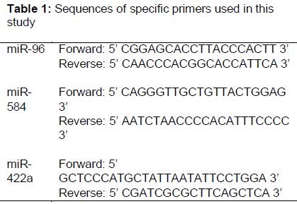
The expression profiling of colon tissue miRNA was performed by Taqman assays. The miRNAs, miR-96, miR-584 and miR-422a, endogenous 5s rRNA primers and probes were purchased from Invitrogen, USA. The sequences of specific primers used in this study are indicated in . The final volume of the PCR mixture was 50 μL and it contained 0.3 μM probe, 0.3 μM of forward and reverse primers, 1X PCR buffer, 0.4 mM dNTPs and Taq DNA polymerase. The thermocycling conditions included initial denaturation at 95 ◦C for 5 min, followed by 35 cycles of 95 ◦C for 20 s, 58 ◦C for 30 s, and 68 ◦C for 40 s. Corresponding negative control without a template was used in all the runs and all the PCR runs were carried out in triplicates. miRNA expression levels were quantified using the ABI Prism 7000 Sequence detection system (Applied Biosystems, USA). The relative expression levels of the miRNAs were calculated using the 2-ΔΔCT method. 5s rRNA was used as an internal control to normalize the data.
Statistical analysis
The differential expression of miRNA between colon cancer samples and the corresponding normal mucosa was assessed by a two-sided Student’s t-test. The relationship between miR-96, miR-584 and miR-422a expression levels and clinicopathological characteristics were analyzed by non-parametric tests. A p-value of < 0.05 was considered statistically significant. All statistical analyses were performed using GraphPad Prism 5 software and Microsoft Excel Version 14.6.5.
Results
Expression levels of miR-96, miR-584 and miR-422a in colon cancer tissues
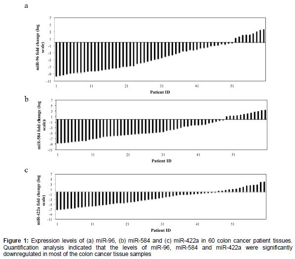
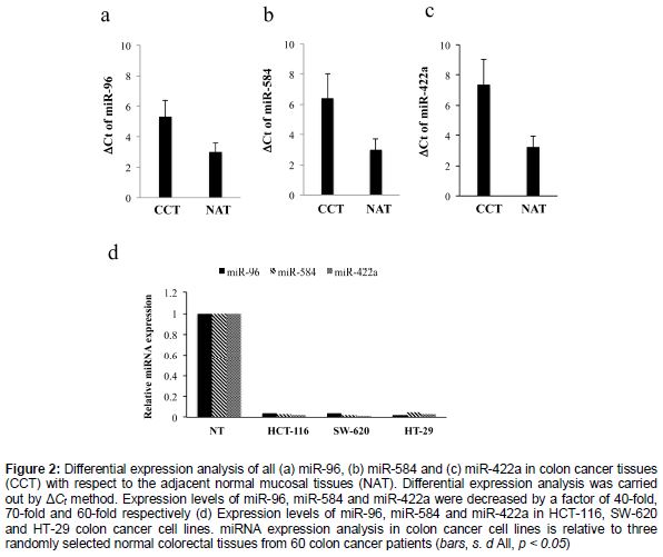
The expression levels of miR-96, miR-584 and miR-422a were analyzed in 60 colon cancer patient tissue samples and their adjacent normal mucosal counterparts using quantitative RT-PCR. Clinical characteristics of the 60 patients involved in this study are summarized in . Quantification analysis indicated that the levels of miR-96, miR-584 and miR-422a were significantly downregulated in most of the colon cancer tissue samples (a, b and c). Next, we went ahead to analyze the differential expression levels of all the three miRNAs with respect to the adjacent normal mucosal tissues. Differential expression analysis using ΔCt method revealed that the expression levels for miR-96 decreased by a factor of 40-fold in 50 out of 60 cancer tissue samples relative to the normal mucosal tissue counterparts (a, p < 0.01). Similarly, the expression levels of miR-584 and miR-422a were decreased by a factor of 70-fold in 55 out of 60 cancer tissue samples, and by a factor of 60-fold in 45 out of 60 cancer tissue samples (b and c, p <0.01).
Expression of miR-96, miR-584 and miR-422a in colon cancer cell lines
We evaluated the expression levels of miR-96, miR-584 and miR-422a in the colon cancer cell lines HCT-116, SW-620 and HT-29. The results indicated significantly lower expression levels of miR-96, miR-584 and miR-422a in all the three colon cancer cell lines compared to the randomized normal mucosal tissue from patients (d).
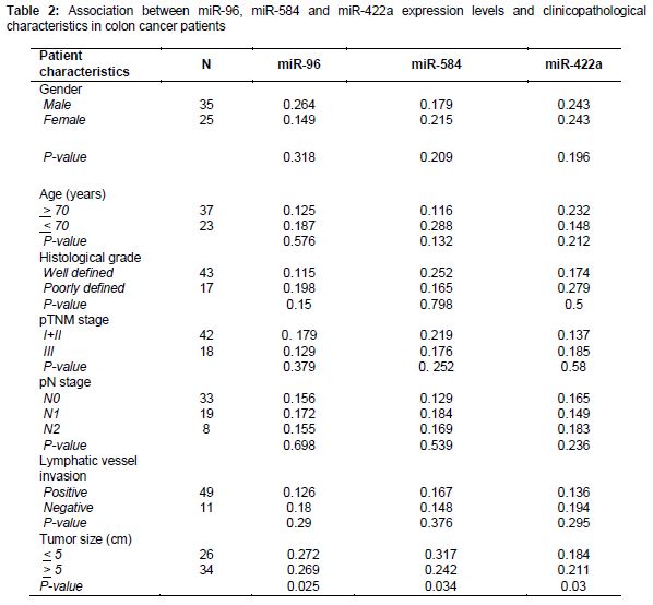
Association between miRNA expression and clinicopathological characteristics of colon cancer patients
We evaluated the association between miR-96, miR-584 and miR-422a expression levels and clinicopathological characteristics in colon cancer patients. No significant correlation could be obtained between low expression levels of miR-96, miR-584 and miR-422a and characteristics such as patients’ gender, age, the tumor and the clinical stages, lymph node metastasis and invasion. However, our correlation analysis suggested a significant correlation between lower expression levels of miR-96, miR-584 and miR-422a and the size of the tumor, irrespective of other clinicopathological characteristics. Our results suggest that, lower expression levels of the aforementioned miRNAs are associated with increased tumor size (, all p < 0.05).
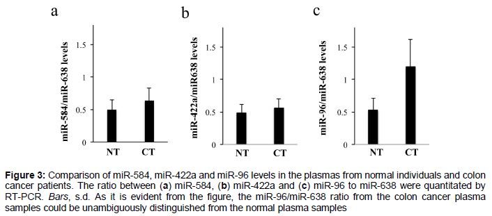
Ratio of miR-96/miR-638 is a potential biomarker for colon cancer
We next wanted to investigate whether the expression levels of miR-96, miR-584 or miR-422a in blood could act as a non-invasive viable biological marker to discriminate colon cancer patients from healthy individuals. We measured the expression levels of miR-96, miR-584 and miR-422a independently in plasma samples from 25 colon cancer patients with respect to the healthy individual samples by quantitative RT-PCR. Recent studies have shown that, since the levels of plasma miR-638 are quite stable, miR-638 can be used as a standard to improve the precision of the analysis [14]. Therefore, we analyzed the ratios of miR-96, miR-584 and miR422a levels to miR-638 levels in the plasma samples from healthy and colon cancer patients. The ratio of plasma levels of miR-584/miR-638 and miR-422a/miR638 from colon cancer patients could not be unambiguously distinguished from the aforementioned ratios from healthy individuals ( a and b). However, the analysis revealed an increased ratio of miR- 96/miR-638 in the plasma samples from the colon cancer patients with respect to the healthy individuals (c). Thus, the levels of miR-96/miR-638 can act as a potential biomarker in the prognosis of colon cancer.
Discussion
The last decade has witnessed advancement in colon cancer research and diagnosis, however it is still one of the deadliest form of cancer with a number of fatalities per year. Since the survival rates of colon cancer patients depend on the early detection of cancer it has become necessary to identify biological markers for a better diagnosis. In the last decade, different classes of molecules have been identified as potential biomarkers for colon cancer and one such intriguing class of molecules is the miRNAs [15]. Large-scale miRnome studies have been carried out in different human solid tumors of breast, colon, pancreas, lung and stomach and identified a large cohort of miRNAs that have been over-expressed or under-expressed [16,17]. Several other studies identified such “miRNA signatures” in cancer and conclusively establish that miRNAs play a critical role in cancer pathogenesis, playing role of either oncogenes or tumor suppressor genes.
Several published literature has provided sufficient evidence that miR-96, miR-584 and miR-422a are often de-regulated in cancer causing pathways. While these miRNAs have been associated with colon cancer and reported previously [18], to the best of our knowledge, a comprehensive validation and characterization of these miRNAs in colon cancer patient tissue samples and cell lines, and the ability of these miRNAs to act as a viable biomarker in the prognosis of colon cancer is still required.
Firstly, the levels of miR-96, miR-584 and miR-422a were assessed in colon cancer tissue and adjacent normal mucosal tissue counterparts using real-time PCR owing to its rapid quantification as well as sensitivity. Significantly lower expression levels of these miRNAs in the cancer tissue compared with the healthy tissue indicated that miR-96, miR-584 and miR-422a are down-regulated in colon cancer. Next, the levels of these miRNAs were assessed in some routinely used colon cancer cell lines.
Similar to the patient tissue samples, HCT-116, SW-620 and HT-29 colon cancer cell lines showed subdued expression levels of these miRNAs. Further, the correlation between low miRNA expression levels and clinicopathological characteristics of colon cancer patients was evaluated. This study indicated an interesting correlation between low expression levels of miR-96, miR-584 and miR-422a with increase in size of the tumor in colon cancer patients.
A similar study has been carried out in the past in 29 colorectal cancer tissues and the study shows the expression of miR-21, -31, -143 and -145 to be altered in CRC and is related to clinicopathological characteristics such as tumor size and lymph node metastasis [19]. Thus, it is evident that miR-96, miR-584 and miR-422a show altered expression levels in colon cancer and it is likely that these miRNAs have the potential as a biomarker in the prognosis of colon cancer.
miRNAs have been shown to have a promising potential as prognostic biomarkers for various types of cancer. Therefore, we investigated the predictive value of miR-96, miR-584 and miR-422a in blood plasma as a viable prognostic biomarker for colon cancer. Quantification of the ratios of miR-584/miR-638 levels and miR-422a/miR638 levels did not yield success, as a clear distinction could not be obtained with respect to healthy individuals. However, we found the ratio of miR-96 to miR-638 to be increased in the plasma samples of colon cancer patients compared to healthy individuals. The distinction was remarkable and thus the levels of miR-96/miR-638 have the potential as a viable non-invasive biomarker for colon cancer. Similar quantification studies were carried out in the plasma samples of hepatocellular carcinoma patients to identify potential biomarkers [14].
Conclusion
This study shows that miR-96, miR-584 and miR-422a are downregulated in colon cancer and are associated with tumor size. The levels of miR-96/miR-638 has the potential of a non-invasive biomarker in colon cancer. Translating this finding into clinical practice would require a thorough and systematic development of diagnostics followed by randomized investigations and validations for demonstrating the efficacy of such non-invasive biomarkers in colon cancer diagnosis.
Declarations
Acknowledgement
References
Archives


News Updates